Field of Science

Comments on Dr. Wolfe-Simon's Response

Preliminary Response to Questions Concerning the Science Article (from F. Wolfe-Simon, Dec. 16, 2010)


(In this post I'm only addressing the specific points made in this Q&A. Magenta numbers identify points I have concerns about. As always, I'm not asking readers to take my word for anything; if you have doubts you should look up the information and check my calculations for yourself.)


Question:
Some people have questioned whether the DNA was sufficiently cleaned by your technique using gel electrophoresis, to separate it from other molecules. Do you feel this is a valid concern?
Answer:
Our DNA extraction and purification protocol begins with washed cells, pelleted from media. These are then subjected to a standard DNA extraction protocol (1), which included multiple phenol chloroform steps to remove impurities, including any unincorporated arsenate (As) (2). After this, the DNA was electrophoresed, further separating the DNA from impurities (3, 4). Any residual As from the media would have been removed by washing the cells prior to extraction (5) and by partitioning into the aqueous phase (6) during the 3 phenol:chloroform steps in the extraction. If As was incorporated into a lipid or protein it would have partitioned into the phenol, phenol:chloroform, or chloroform fractions (7). Additionally, DNA extracted in this manner on other samples was also successfully used in further analyses, including PCR (8), that require highly purified DNA (9).
The arsenic measured by NanoSIMS in the gel band is consistent with our other measurements and another line of evidence.
Our radiolabeled 73AsO43- experiment showed that of the total radiolabel associated with the cell pellet 11.0 % ± 0.1 % was associated with the DNA/RNA fraction (10). This indicated that we should expect some arsenate of the total pool associated with the nucleic acids (11). To interpret these data, we coupled our interpretation with our EXAFS evidence suggesting that intracellular arsenic was As(V) bound to C (12), and was not free in solution as an ion. This suggests the As is in, an organic molecule with bond distances consistent with a chemical environment analogous to phosphate (Figs. 3A, S3 "bond lengths" table). Further supporting our interpretation of the previous mentioned two analyses, we used a third line of evidence from NanoSIMS, a completely different technique from the other two. We find elemental arsenic (as measured by NanoSIMS) associated with the gel band that is more than two times the background in the gel (13). Based on the above discussion, we do not feel this is a valid concern.

My concerns:
(1) The DNA extraction procedure included only some of the components of a standard DNA extraction protocol.  First, only a single ethanol precipitation was done, whereas getting relatively pure DNA requires at least two rounds of extraction and precipitation.  Second, the pellets were not washed, so that unincorporated arsenate (or phosphate) present in the aqueous fractions may have been precipitated with the DNA, and may also have been present in the alcohol supernatants contaminating the pellets.  Third, no column clean-up step was done.
(2) This statement implies that unincorporated arsenate in the cell lysate would partition into the phenol and chloroform.  This seems a priori improbable, as arsenate is very soluble in water.  No controls were done to find out how unincorporated arsenate or phosphate would partition in these extractions.
(3) Gel electrophoresis can remove impurities but it is not guaranteed to do so.  Any impurities that migrate at a similar rate to the DNA, or are electrostatically associated with it, will be present in the gel slice.  Any impurities that diffuse into the gel buffer may become distributed throughout the gel.  Any impurities already present present in the agarose or gel buffer will also be in the gel slices.  These concerns are strengthened by the failure to purify the DNA away from the gel slice (see (13) below).
(4) No control was performed for non-covalent association of arsenate (or phosphate) with DNA.  In another control extraction, arsenate and lysis solution should have been mixed with previously purified DNA from E. coli or other phosphate-grown cells, to see if any arsenic co-purified with the DNA. 
(5) No control was performed for the effectiveness of this washing.  E. coli or other cells grown in the absence of arsenate should have been mixed with the +As/-P medium and then subject to the same washing and extraction steps.
(6) Here arsenate is predicted to partition into the aqueous phase.  Is this the same arsenate that partitioned into the phenol and chloroform in (2) above?
(7) In the ICP-MS analysis presented in Table S1, almost all the arsenic did partition into the phenol phase, and almost as much arsenic was present in the phenol fraction of the phosphate-grown cell (4725 vs 3683 ppb).  In fact, the aqueous phase of arsenate-grown cells contained no detectable arsenic at all, even though this is the fraction from which the arsenic-containing DNA was precipitated. 
(8) The online Methods say that DNA from all growth conditions, worked fine in the PCR reactions used for the phylogenetic analysis.  This strongly suggests that the DNA from arsenate-grown cells has a normal phosphorus backbone.  The polymerases used for PCR have very high fidelity and would not tolerate substitution of arsenic for phosphorus.
(9) PCR does not require purified DNA; it even works very well on whole-cell lysates.
(10) See point (7)
(11) But this pool is expected to contain all of the water-soluble constituents of the cell. The elemental analysis report that the arsenic was bonded to carbon doesn't mean that it is bonded to DNA.  And most of this arsenic partitioned into the phenol phase - is it thought to be lipid?  If so, maybe the arsenic is bonded to C in lipids.  
(12) Wait!  IANAC (I am not a chemist), but if the 'intracellular arsenic was As(V) bound to C' then it couldn't be arsenic incorporated into DNA or RNA, as it would then have to be bound to O in DNA's diester backbone.
(13) Let's think more about the arsenic in the gel bands.  The whole gel slices were assayed (the DNA was not purified away from the agarose); since the gel is 1% agarose and a gel slice is unlikely to weigh less than 100 mg, each slice would contain at least 1 mg of agarose. The DNA bands in Fig. 2A are unlikely to contain more than 1 µg of DNA, probably less for arsenate-grown DNA in lane 2).  Thus we can generously assume that 99.9% of the carbon in each DNA sample came from the agarose, and no more than 0.1% from the DNA.  According to the figure legend and the numbers at the bottoms of the gel lanes the arsenate-grown sample had 13.4 atoms of arsenic per 10^6 atoms of carbon.  This is 13.4 arsenic atoms per 1000 DNA carbons.  Since A, G and T nucleotides contains 10 carbons  and C has 9, this is 13.4 arsenic atoms per 102.6 nucleotides, or about 26 per 100 base pairs.  That's quite a lot of arsenic.  Even more surprising, the phosphate-grown sample had 6.9 arsenics per 10^6 carbons, which would be about 14 arsenics per 100 bp.  The gel blank had even more arsenic, and three times as much phosphorus.  This strongly suggests that the gel was contaminated with both arsenic and phosphorus, perhaps introduced with the DNA samples.  Until such contamination can be ruled out, the two-fold higher arsenic concentration and three-fold lower phosphorus concentration associated with the arsenic-grown DNA sample cannot be seen as significant.

Question:
Others have argued that arsenate-linked DNA should have quickly fallen apart when exposed to water. Could you address this?
Answer:
We are not aware of any studies that address arsenate bound in long chain polyesters or nucleotide di- or tri-esters of arsenate, which would be directly relevant to our study. Published studies have shown that simple arsenic esters have much higher hydrolysis rates than phosphate esters (1-3). The experiments published to date have specifically looked at the exchange or hydrolysis of alkyl tri-esters of arsenate [Eqn. 1] and alkyl di-esters of arsenite [Eqn. 2]:
OAs(OR)3 + H2O → OAs(OH)(OR)2+ ROH [1]
OAs(OH)(OR)2 + H2O → OAs(OH)2(OR) + ROH [2]
where R = methyl, ethyl, n-pentyl and isopropyl. Reference 2 demonstrated that the hydrolysis rates for these simple alkyl triesters of arsenate decreased with increasing carbon chain length (complexity) of the alkyl substituent (methyl > ethyl > n-pentyl > isopropyl) (14). No work has been done on the hydrolysis rates of arsenate-linked nucleotides or other biologically relevant moieties.
If the hydrolytic rate trend reported in Ref. 2 continues to larger-weight organics, such as those found in biomolecules, it is conceivable that arsenate-linked biopolymers might be more resistant to hydrolysis than previously thought (15). The small model compounds investigated in Refs. 1-3 are relatively flexible and can easily adopt the ideal geometry for water to attack the arseno-ester bond (16). Arsenate esters of large, bio-molecules, however, are likely to be more sterically hindered leading to slower rates of hydrolysis (17).
This type of steric constraint on reaction rate accounts for the wide range of rates seen in the behavior of some phosphate linked nucleotides. In small ribozymes, the phophodiester linkages at the site of catalysis can be hydrolyzed on the order of tens of seconds (with a chemical rate of 1 s-1). This rate enhancement is achieved by orienting the linkage for in-line attack by a nucleophile (an adjacent 2' hydroxyl group). Moreover, the autodegradation patterns are consistent with specific base composition. On the other hand, the hydrolysis rates for phosphodiester bonds in A form duplexes of RNA are many orders of magnitude slower, because these linkages cannot access easily the geometry necessary for hydrolysis.
The rates in DNA may be much slower than model compounds because of the geometrical constraints imposed upon the backbone by the helix (18).
The kinetics of the hydrolysis of arsenate-linked biopolymers is clearly an area where more research is warranted.

My concerns:
(14) Again IANAC.  But note that these are hydrocarbons and thus quite hydrophobic, especially the pentyl chain (5 carbons) and the isopropyl chain (3 branching carbons).  Attaching three of these by ester bonds to the arsenate effectively surrounds the bonds with hydrophobic shells that exclude water.  Thus it's not surprising that the hydrolysis reaction occurs less often.
(15) But if the increased stability described in Reference 2 (yes, I looked at this paper, and I'm trying to get hold of the 1870 reference too) is due to increasing hydrophobicity of the ester bonds' environment, then the effect will not extrapolate to long hydrophilic biological molecules such as DNA.
(16) Not if they're surrounded by a hydrophobic shell, and probably also associating with each other to reduce the shell's exposure to the aqueous solvent.
(17) DNA has its backbone on the outside of the double helix, and the entire molecule is quite hydrophilic.  
(18) Assuming the overall structure of arsenic bonds in DNA is like that of phosphorus bonds in DNA, shouldn't any stability-enhancing geometrical constraints of the DNA structure be experienced by both phosphorus and arsenic bonds?  We would then still expect arsenic bonds in DNA to be 100-fold less stable than phosphate bonds. 

Question:
Is it possible that salts in your growth media could have provided enough trace phosphorus to sustain the bacteria?
Answer:
The data and sample labeling in Table S1 has caused some confusion. To clarify, for every experiment, a single batch of artificial Mono Lake water was made with the following formulation: AML60 salts, no P, no As, no glucose, no vitamins. Table S1 shows examples of ICPMS measurements of elemental phosphorus (~3 µM) and arsenate made on this formulation prior to any further additions (19, 20). Then we added glucose and vitamins for all three treatments and either As for the +As treatments or P for the +P treatments. The P measurements made on the medium after the addition of sucrose and vitamins and after addition of As were also ~3 µM in this batch. Therefore, it was clear that any P impurity that was measured (~3 µM, this was the high range) came in with the major salts, and that all experiments contain identical P background (including any P brought in with the culture inocula).

In the Science paper, we show data from one experiment of many replicated experiments that demonstrates no growth of cells in media without added arsenate or phosphate (Figure 1). These data clearly demonstrate that strain GFAJ-1 was unable to utilize the 3µM P to support further growth in the absence of arsenate (21). Moreover, the intracellular P content determined for the +As/-P grown cells was not enough to support the full requirement of P for cellular function (22).
Note on culturing: All experiments were initiated with inocula from sustained +As/-P conditions. Prior to the experiments, the cells had been grown long term, for multiple generations from a single colony grown on solid media with no added phosphate. Before this, they were grown as an enrichment for more than 10 transfers and always into new medium that was +As/-P. We therefore feel that there is not significant carry-over of P. We also argue that there would not have been enough cellular P to support additional growth based on an internal recycling pool of P (23).


My concerns:
(19) The two batches of AML60 salts assayed contained 3.7 and <0.3 µM P, and the single batch of cell wash solution contained 7.4 µM P.  Given this variability, the similarity of the two batches of -P/+As medium doesn't inspire much confidence.
(20) Was no effort made to identify and eliminate the source(s) of this contamination?
(21) Agreed, with the proviso that the media be tested and shown to be identical except for the phosphate and arsenate.  But this wouldn't mean that arsenic replaced phosphorus in any biological molecules in GFAJ cells, just that the cells needed arsenate for something.
(22) Did this calculation take into account the very high carbon content of the poly-hydroxybutyrate granules in these cells?  PHB can account for up to 90% of the dry weight of phosphate-starved cells, and its carbon will skew estimates of C:P ratios.
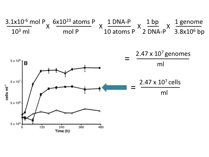
(23) This assertion is not supported here by any evidence, and it is contradicted by the Makino et al. (2003) reference cited by the Science paper (ref. 13). These authors found that 10% of the P in phosphate-limited E. coli is in the DNA and the rest is in RNA and other cellular components.  Using this value and your own estimate of genome size (3.8 Mbp), ~3 µM P is sufficient to account for the observed growth in the -P/+As medium. Here's the calculation:  


The FedEx saga continues

Regular readers (all 5 of you) may remember last week's unsuccessful attempt to ship some of our bacteria to London.  FedEx returned our package to us because we hadn't met all their stringent requirements for infections bacteria.

This afternoon we went back to Shipping and Receiving with all the new correct FedEx paperwork and our 24 vials of bacterial cells.  The vials were in a plastic box, in a special leakproof container, in this container's specially labeled cardboard carton, in a big styrofoam container for the dry ice, in a very big cardboard box labeled OVERPACK.

Only to learn that Shipping and Receiving had run out of dry ice! 

It was too late to rush over to Chemistry Stores to get dry ice, as we'd miss the FedEx pickup time.  I briefly considered finding an open-late FedEx office and hand-delivering the package to them.  This would have involved sending someone over to Chemistry Stores to get the dry ice while I rode my bike home (5 miles) to get my car.  But then I remembered that I'm probably not authorized to even touch this hazardous-goods shipment, much less drive it around town.

Shipping and Receiving won't have dry ice until the day after tomorrow, so tomorrow one of us will go over to Chemistry Stores and get the dry ice we need, and we'll try yet again to get the shipment on its way to London.

How to harness distributed discussion of research papers

In this post I'm going to elaborate on a suggestion I saw a few days ago, in an article discussing the role of post-publication commentary in science.  (And yes, I'm searching for the source of this idea - if any reader remembers whose it was, please point me to it.)

In modern but pre-internet days, researchers did the research, wrote the paper, submitted it to peer review, made changes, and published it.  Other researchers them evaluated this information, using to guide their own work, and discussed its strengths and weaknesses when they cited it in their own papers. 

Published papers were also discussed less formally with colleagues, both before and after publication,  face-to-face and by mail and phone, and in journal-club presentations and seminars.  The ideas from these discussions were incorporated into the formal papers drawing on this work, but they weren't available to anyone but the direct participants.

Now that we're all on line, published papers are also being discussed more publicly, in blogs and other places.  Such discussions are extraordinarily valuable for the progress of science - they're written public evaluations, drawn from a wide range of expertise, and usually greatly enriched by comments from and links other researchers.  But these pages are all over the place, and finding them requires a lot of active searching.

The Research Blogging site is helping with this problem, by aggregating blog posts that discuss individual research papers.  But they can only link to posts that actively insert their code, and so miss quite a lot of the public commentary.  So far the journals don't link their papers to this site, so readers who go looking for the paper don't usually think to also check Research Blogging.

A few forward-thinking online journals (PLoS and BMC groups, I'm talking about you) provide their own Comments thread for each paper, so other researchers can provide informal but public feedback .  But the researchers don't use these, saying that they don't feel comfortable doing this publicly, or that they don't like the bother of having to register and log on.  I know that's true for me, thought I don't know why - I'll happily blog about a paper I've read, but I almost never post comments on its official Comments page.

Sites like The Third Reviewer have tried to solve this by providing journal-independent sites where researchers can post comments about papers.  But we won't use these either -the massive wave of discussion about the Wolfe-Simon paper on arsenic bacteria let to exactly zero comments on The Third Reviewer.

Most journals already provide, with each paper they've published, a list of links to the more recent papers that cite it.  The suggestion I really liked was that the journals should also aggregate the informal commentary, by providing a separate list of links to ALL the web pages that have link to the paper.  Journals could then stop fighting our unwillingness to post comments centrally, and just use our distributed posts to add value to the papers they publish.

I don't think this would be very difficult; lots of sites already have a 'Who links here?' feature, and I think both Google and Yahoo searches can be restricted to sites containing a specific url.  The journals could include an explicit disclaimer that the journal in no way vouches for the value or creditability of the information in these links.  A few bloggers might have to become a bit more circumspect (probably not a bad thing), and any blogger who didn't want their post linked to the paper could provide a citation to the paper but not link to it.

Jon Eisen,  do you think this could work?  If PLoS leads, I bet the others will follow.

An apologetic email

Note added Dec. 13:  The author of the ABC article has modified it (even though he had accurately quoted what I'd said to him), and he now also quotes from this email.

Dear Dr. Wolfe-Simon,

I'm emailing you to apologize for quotes from me that have just appeared on the ABC News website.  I wasn't misquoted, but some of the things I said in a phone interview yesterday morning came across more harshly than I had intended.

I told the interviewer that, even though I think your conclusions were wrong, I sympathize with the difficult position you're in (I've spent about 20 years championing a hypothesis that almost everyone thinks is wrong).  I also said that what matters in science isn't whether we make mistakes (we all do) but how we deal with them, and that I think you're handling the situation well.

I feel particularly bad about the 'not calm and confident' quote, because in fact your press conference was very well done.  I meant this statement to only emphasize that women in science know that they're being judged harshly, but instead I came across as someone doing precisely that.

Sorry,

Rosie Redfield

p.s. to everyone else:  I don't want this post to become a place to debate speaking styles so I'm going to close comments (or delete them if I can't figure out how to close them).  My apologies to the four people who've already commented, as I'm about to delete your comments.

Back to blogging as usual

But I can't resist first posting this fabulous photo-mashup of a face-off between myself and Felisa Wolfe-Simon (from Gizmodo):

Now, where was I?  Last week's transformation experiment worked quite well.  I didn't get nearly as many NovR and NalR transformants as I had expected, and some of the plates had contaminants, but I ended up with five independent pools containing between 5,000 and 50,000 independent clones each.  So I diluted each pool to about 3 x 10^9 cells per ml, added glycerol, and froze four aliquots of each pool in nicely labelled tubes, all ready to FedEx to London (England, not Ontario) on Monday.

Well, it's Thursday, and the cells are still in our freezer.  Actually they're back in our freezer, because we shipped them out yesterday afternoon, thinking we had met all the requirements, and this morning FedEx brought them back.   (But we still have to pay them for this on-shipment!)

The problem is that these are infectious bacteria, and the shipping agencies enforce very strict regulations about packaging and labelling.  I think some central agency (government) must make the regulations, and packages must be prepared for shipping by a person who's taken a special training course in transport of hazardous goods.  I probably should have realized this, but I didn't, and we had a really hard time finding the information we needed.  (We haven't had to do this in the past - we've usually just sent people DNA rather than cells.)

Our Shipping and Receiving office originally told the RA to talk to Health and Safety, and Health and Safety didn't return the RA's call or her email.  So on Tuesday I talked to FedEx, and then I called up Health and Safety and found out that (1) the shipper had to have taken a course; (2) it's not the kind of course you can just take in an hour online; (3) a technician in our building had taken the course and might help us.  Then she told us that the manager of Shipping and Receiving had the training we needed, and sure enough he did. But it was too late to do it then.

He was busy Wednesday morning, but yesterday afternoon we took the frozen cells inside the special o-ring sealed plastic container inside the specially labelled cardboard box (we had received the container and box when someone else had shipped cells to us) inside the big styrofoam box.  He got us the dry ice, checked all our paperwork and labels, signed the forms, and put his cell phone as the emergency 24-hr contact number.  The RA had already set up the shipment online and printed out the waybill and commercial invoice (3 copies) and the Dangerous Shipment declaration (3 copies, each printed in colour on the lab next door's printer).  She taped the styrofoam box shut and put in the special FedEx pickup place, and we both heaved sighs of relief.

But this morning the box came back.  Once it had reached FedEx's central clearing house they'd gone over it with their 900-point checklist (I exaggerate only slightly) for hazardous-goods shipments, and it had failed.  Not just one point - there were Xs in about ten of the boxes.  We had forgotten to write the weight of the dry ice on the form.  The shipping guy had forgot to sign one of the forms in one of the places, and to resign at somewhere a change had been made.  Our styrofoam box needed to be inside a cardboard box, which must be labelled "OVERPACK".   The Dangerous Shipment form must describe the contents with very precise wording.  And it must not be completed by hand - of course we don't have a typewriter, and it's on a pdf form that can be typed into but not saved, so we'll ask the lab next door to let us borrow one of their computers as well as their colour printer - we can't just complete the form on one of our computers and send it to their printer because we're in different departments and thus our computers are on different networks.

Because the shipment will probably take longer than overnight to get to London, we didn't want to send it on a Thursday or Friday and risk having it sit around getting warm all weekend.  So we're getting everything ready again to send it on Monday.

Interested in doing research in my lab?

I'm taking advantage of this barrage of visitors to spread the word that my research group has openings for both a graduate student (M.Sc. or Ph.D.) and a postdoctoral fellow.  We investigate the molecular biology and evolution of bacteria DNA uptake; you can read more about us at our home page, and find the details of our current research plans in our latest grant proposals.

My Letter to Science

(If you're looking for the long post I wrote on Saturday, the one that started the controversy about the Wolfe-Simon arsenic bacteria paper by describing all the problems I found, it's here.)

Below is the text of my formal Letter to Science about the Wolfe-Simon paper.  Letter submissions are supposed to be limited to 300 words (this is a bit over at 371), so I'm only bringing up the issues concerning contamination.  This is an improved version that incorporates many of the suggestions provided in the comments below.  So some of the comments (in the first 10-15) won't make sense any more.

Because this paper has LOTS of other problems, it would be great if many other researchers could also submit Letters and Technical Comments (limit of 1000 words, peer-reviewed).  Here's a link to the Instructions for Authors entry page.  I won't mind if your Letter gets accepted and mine doesn't.

Here's mine:


Wolfe-Simon et al. (1) meticulously eliminated contamination of the reagents and equipment used in their elemental analyses, but they made much less effort to eliminate contamination in their biological samples.

The reagents used for the culture media were not pure.  The 3.1 µM PO4 contaminating the As+/P- medium provided enough P for all of the cell growth seen in this medium, using the authors’ estimate of 7.5x10⁶ atoms of P per genome and the generous assumption that phosphate-starved cells use 90% of their P for molecules other than DNA (2).  This calculation (not done by the authors) obviates their hypothesis that the cells could only grow by replacing P with As.

An independent contamination problem is the omission of standard DNA purification steps when testing for As in DNA (2).  Contamination is typical in DNA/RNA pellets produced by ethanol precipitation of the aqueous phases from phenol:chloroform extractions.  This is partly because this fraction contains most of the small molecules from the cytoplasm (contrary to the authors’ assertion), which are often less soluble in 70% ethanol than in water.  Pellets are also typically contaminated with small amounts of the ethanol supernatant.  Yet the usual step of washing the pellets was omitted, and the dried pellets were simply resuspended in water and loaded on an agarose gel.

Most surprisingly, the chromosomal DNA fractions (boxed in Fig. 2A) were not purified from the gel slices (a standard ten-minute procedure).  Instead the authors simply dried the gel slices and assayed them.  Not only does this bring in any contaminants present in the gel, but since each gel slice would have contained at least 1 mg of agarose (100 mg of 1% agarose gel), and each DNA band no more than 1 µg of DNA, at least 99.9% of the carbon in these samples would have come from the agarose, not the DNA.  No correction can be made for the agarose-derived C because the actual amounts of DNA and agarose are not known.  Omission of the gel-removal step for these critical samples is surprising because the authors did use it in preparing the rDNA fragments they sequenced for their phylogenetic analysis.

1.      Wolfe-Simon F, Blum JS, Kulp TR, Gordon GW, Hoeft SE, Pett-Ridge J, Stolz JF, Webb SM, Weber PK, Davies PC, Anbar AD, & Oremland RS (2010). Science Express. PMID: 21127214
2.      W. Makino, J. Cotner, R. Sterner, J. Elser, Funct Ecol 17,121 (2003).
3.      J. Sambrook, D. W. Russell.  Molecular Cloning, A Laboratory Manual. 3rd Ed.  Cold Spring Harbor Press, New York 2001.

Arsenic-associated bacteria (NASA's claims)

ResearchBlogging.org

Wolfe-Simon F, Blum JS, Kulp TR, Gordon GW, Hoeft SE, Pett-Ridge J, Stolz JF, Webb SM, Weber PK, Davies PC, Anbar AD, & Oremland RS (2010). A Bacterium That Can Grow by Using Arsenic Instead of Phosphorus. Science (New York, N.Y.) PMID: 21127214

Note to visitors in 2012:  We've just submitted a manuscript to Science reporting the results of our unsuccessful attempt to replicate the key findings of this work.  The manuscript will be publicly available on the arXiv server beginning Feb. 1 2012.


Newer note to new readers:  See also my new (Dec. 16) critique of the authors' response to these and similar criticisms.)

Note to new readers:  I wrote this post on Saturday Dec. 4, mainly to clarify my own thinking.  I didn't expect anyone other than a few researchers to ever read it.  Since then I've made a few minor corrections and clarifications (typos, decimal places, cells not cfu), but I haven't changed anything significant.  Please read the comments - they contain a lot of good scientific thinking by other researchers.

Here's a detailed review of the new paper from NASA claiming to have isolated a bacterium that substitutes arsenic for phosphorus on its macromolecules and metabolites.  (Wolfe-Simon et al. 2010, A Bacterium That Can Grow by Using Arsenic Instead of Phosphorus.)  NASA's shameful analysis of the alleged bacteria in the Mars meteorite made me very suspicious of their microbiology, an attitude that's only strengthened by my reading of this paper.  Basically, it doesn't present ANY convincing evidence that arsenic has been incorporated into DNA (or any other biological molecule).

What did the authors actually do?  They took sediment from Mono Lake in California, a very salty and alkaline lake containing 88 mg of phosphate and 17 mg of arsenic per liter.  They put the sediment into a similarly alkaline and hypersaline defined medium containing 10 mM glucose as a carbon source, 0.8 mM NH4SO4 as a nitrogen and sulfur source, and a full assortment of the vitamins and trace minerals that might be needed for bacterial growth.  Although this basic medium had no added phosphate or arsenate, contamination of the ingredients caused it to contain about 3 µM phosphate (PO4) and about 0.3 µM arsenate (AsO4).  For bacterial growth it was supplemented with arsenate or phosphate at various concentrations.

The interesting results came from sediment originally diluted into medium supplemented with the highest arsenate concentration they initially tried (5 mM) but no phosphate.  Over the course of several months they did seven tenfold dilutions; in the sixth one they saw a gradual turbidity increase suggesting that bacteria were growing at a rate of about 0.1 per day.  I think this means that the bacteria were doubling about every 10 days (no, every 7 days - corrected by an anonymous commenter).

After one more tenfold dilution they put some of the culture onto an agar plate made with the same medium; at least one colony grew, which they then inoculated into the same defined medium with 5 mM arsenate.  They gradually increased the arsenate to 40 mM (Mono Lake water contains 200 µM arsenate).  Descendants of these cells eventually grew in 40 mM arsenate, with about one doubling every two days.  They grew faster if the arsenate was replaced by1.5 mM phosphate but grew only about threefold if neither supplement was provided (Fig. 1 A and B, below).  The authors misleadingly claim that the cells didn't grow at all with no supplements.

In Fig. 1 (below), the correspondence between OD600 (Fig. 1 A) and cells (Fig. 1 B) is not good.  Although the lines in the two graphs have similar proportions, OD600 is plotted on a linear scale and cells/ml on a log scale (is this a shabby trick to increase their superficial similarity?).  OD600 in arsenate medium was almost as high as that in phosphate medium, but the number of cells was at least tenfold lower.  And the OD in arsenate continued to increase for many days after the cells has leveled off.  I suspect most of the continuing growth was just compensating for cell death.  It would be interesting to test whether the cells were scavenging phosphate from their dead siblings.  (A researcher in my lab had a better explanation - I've put it in the Comments below.)


The authors never calculated whether the amount of growth they saw in the arsenate-only medium (2-3 x 10^7 cfu/ml) could be supported by the phosphate in this medium (or maybe they did but they didn't like the result).  For simplicity I'll start by assuming that a phosphorus-starved cell uses half of its phosphorus for DNA and the rest for RNA and other molecules, and that the genome is 5x10^6 bp.  Each cell then needs 1x10^7 atoms of phosphorus for DNA, and 2x10^7 for everything.  The medium is 3.1 µM phosphate, which is 3.1x10^-6 moles per liter.  Mutiply by Avogadro's number (6.02x10^23 atoms per mole) and we have 1.9x10^18 atoms of phosphorus per liter, or 1.9x10^15 per ml.  Divide by the phosphorus requirement of each cell (2x10^7) and we get 9.5 x 10^7 cells per ml.  This value is just comfortably larger than the observed final density, suggesting that, although these bacteria grow poorly in the absence of arsenate, in its presence their growth is limited by phosphate. (Note:  This calculation originally dropped a decimal point.  I've changed it a bit and corrected the error.)

Under the microscope the bacteria grown with arsenate and no added phosphate (Fig. 1 C) look like plump little corn kernels, about 1 µm across and 2 µm long.  They contain many structures (Fig. 1 E) which the authors think may be granules of the wax-like carbon/energy storage material polyhydroxybutyrate (PHB).  Many bacterial cells produce BHP when their carbon/energy supply is good but other nutrients needed for growth are in short supply.  Cells grown with phosphate and no added arsenate are thinner and lack the granules (Fig. 1 D).  The authors used 16S rRNA sequencing to identify this bacterium as belonging to the genus Halomonas, a member of the gammaproteobacterial order Oceanospirillales.  Members of this group are diverse but not known to have any uniquely dramatic features.

According to an interview with the first author, this research was motivated by a desire to show that organisms could use arsenic in place of phosphorus.  The two atoms have very similar chemical properties, but bonds with arsenic are known to be much less stable than those with phosphate, so most researchers think that biological molecules containing arsenic rather than phosphorus would be too unstable to support life.  Thus the authors wanted to show that the bacteria had incorporated the arsenic in places where phosphorus would normally be found.  They used several methods, each involving a low-tech preparation of cell material and a high-tech identification of the atoms present in the material.
 
First they collected the bacteria by centrifugation, washed them well, and precisely measured the fraction of arsenic and phosphorus (as ppb dry weight, Tables 1 and S1).  Cells given only the arsenate supplement contained about 10-fold more arsenic than phosphorus (0.2% arsenic and 0.02% phosphorus) and cells given only the phosphate supplement had 0.5% phosphorus and only 0.001%  arsenic.

The authors argue that the arsenate-grown cells don't contain enough phosphorus to support life.  They say that typical heterotrophic bacteria require 1-3% P to support life, but this isn't true.  These numbers are just the amounts found in E. coli cells grown in medium with abundant phosphate.   They are very unlikely to apply to bacteria growing very slowly under phosphate limitation, and aren't even true of their own phosphate-grown bacteria (0.5% P).  The large amount of PHB in the arsenate-grown cells would have skewed this comparison - PHB granules are mainly carbon with no water, and in other species can be as much as 90% of the dry weight of the cells.  Thus their presence only in arsenate-grown cells could depress these cells' apparent phosphate concentration by as much as 10-fold.

The authors then grew some cells with radioactive arsenate (73-As) and no added phosphate, washed and dissolved them, and used extraction with phenol and phenol:chloroform to separate the major macromolecules.  The protein fraction at the interface between the organic and aqueous phases had about 10% of the arsenic label but, because the interface material is typically contaminated with liquid from the aqueous phase, this is not good evidence that the cells' protein contained covalently-bound arsenate in place of phosphorus.  About 75% of the arsenic label was in the  aqueous (upper) fraction.  The authors describe this fraction as DNA/RNA, but it also contains most of the small water-soluble molecules of the cell, so its high arsenic content is not evidence that the DNA and RNA contain arsenic in place of phosphorus.  The authors use very indirect evidence to argue that the distribution of arsenic mirrors that expected for phosphate, but this argument depends on so many assumptions that it should be ignored.

(They also measured the absolute amounts of arsenic and phosphorus in the supernatant fraction - surprisingly, no arsenic (<20 ppb) was detected in the fraction from arsenate-supplemented cells, although the fraction from phosphate-grown cells had 118 ppb!  See Table S1.)

They especially wanted to show that the cells' DNA contained arsenic in place of phosphorus, so they gel-purified chromosomal DNA from cells grown with arsenate (lane 2) or with phosphate (lane 3), and measured the ratio of arsenic to carbon by mass spectrometry.  The numbers at the bottom give these ratios (the legend says 'multiplied by 10^-6 but they surely mean 'multiplied by 10^6'). 


As expected, this ratio was very low for the phosphate-grown cells (6.9x10^-6), but it was only twofold higher for the arsenate-grown cells (13.4x10^-6).  Normal DNA has one phosphorus atom for each ten carbons (P:C = 10^-1), so the arsenate-grown ratio is only about one arsenic atom per 10,000 phosphorus atoms (i.e. one per 5 kb of double-stranded DNA).  A 2x10^6 bp genome would contain 4x10^6 atoms of phosphorus, so if all this arsenate was really covalently in the DNA, each genome would only contain about 400 atoms of arsenic.  And a phosphate-grown genome would contain 200!

Could 400 atoms of arsenate per genome be due to carryover of the arsenate in the phenol-chloroform supernatant rather than to covalent incorporation of As in DNA?   The Methods describes a standard ethanol precipitation with no washing (and no column purification which would have included washing), so I think some arsenate could easily have been carried over with the DNA, especially if it is not very soluble in 70% ethanol.  Would this arsenate have left the DNA during the gel purification?  Maybe not - the methods don't say that the DNA was purified away from the agarose gel matrix before being analyzed.  This step is certainly standard, but if it was omitted then any contaminating arsenic might have been carried over into the elemental analysis.

Failure to purify the DNA away from the agarose would also compromise their elemental analysis in other ways, since much of the carbon in the purified 'DNA' would have been from the agarose.  The authors did do the same elemental analysis on a gel slice with no DNA in it, a control that only makes sense if they didn't purify the DNA.  Not purifying away the gel might affect the arsenate-grown DNA more because the band contains less DNA; this would explain why this excised DNA has 3.5-fold lower ratio of phosphorus to carbon than the phosphate-grown DNA, a difference that is certainly not explained by its very low arsenic content.)

(Might they have not presented assays using properly purified (washed) DNA because these turned out to not have any arsenic?  Am I just paranoid?)

Finally, the authors examined the chemical environment (neighbouring atoms and bonds) of the arsenic in the cells using synchrotron X-ray studies.  This is over my head, but they seem to be trying to interpret the signal as indicating that the environment of the arsenic is similar to that of phosphorus in normal DNA.  But the cellular arsenic being in DNA can't be the explanation, because their DNA analysis indicated that very little of the cellular arsenic purifies with the DNA.  The cells contained 0.19% arsenic (1.9x10^6 ppb), but the DNA only contained 27 ppb arsenic.

Bottom line:  Lots of flim-flam, but very little reliable information.  The mass spec measurements may be very well done (I lack expertise here), but their value is severely compromised by the poor quality of the inputs.  If this data was presented by a PhD student at their committee meeting, I'd send them back to the bench to do more cleanup and controls.

There's a difference between controls done to genuinely test your hypothesis and those done when you just want to show that your hypothesis is true.  The authors have done some of the latter, but not the former.  They should have mixed pregrown E. coli or other cells with the arsenate supplemented medium and then done the same purifications.  They should have thoroughly washed their DNA preps (a column cleanup is ridiculously easy), and maybe incubated it with phosphate buffer to displace any associated arsenate before doing the elemental analysis.  They should have mixed E. coli DNA with arsenate and then gel-purified it.  They should have tested whether their arsenic-containing DNA could be used as a template by normal DNA polymerases.  They should have noticed all the discrepancies in their data and done experiments to find the causes.

I don't know whether the authors are just bad scientists or whether they're unscrupulously pushing NASA's 'There's life in outer space!' agenda.  I hesitate to blame the reviewers, as their objections are likely to have been overruled by Science's editors in their eagerness to score such a high-impact publication.

Planning a transformation experiment

I've promised to transform our lab strain (Rd) with chromosomal DNA from another strain, and send the pooled transformants to another lab for analysis.  Here I need to plan what I'll do.

Because most of the cells in our competent-cell props aren't actually competent, I'm going to transform the cells with a mixture of the other strain's DNA and a short fragment carrying an antibiotic resistance allele.  By selecting for this allele I'll make sure that all the cells I send actually did take up DNA.

I have two fragments I can use; both are about 2.5 kb long, produced by PCR from genomic DNA (the postdoc is making them for me right now).  One carries novobiocin resistance and the other carries nalidixic acid resistance.  I think I should use both, in separate transformations, as this will control for the slight possibility that the unmapped gene they're looking for is in the selected fragment.

How much of each DNA should I use?  I want a saturating amount of the chromosomal DNA, which I have already prepared; 1 µg in a 1 ml transformation should be fine. I should use a lot less of the fragment, because I want most of the DNA the cells take up to be chromosomal.  But I need to use a substantial amount, as I want to have at least thousands and preferably hundreds of thousands of transformants.  Transformation frequencies with pure fragments are typically high, with saturation reached at about 20 ng (I think).  If the transformantion frequency with this fragment was 1%, I would expect to get about 10^7 transformants from 1 ml (10^9) cells, so using 1-10 ng of fragment DNA should be lots.

I can do several controls to check how much of the chromosomal DNA the antibiotic-resistant transformants had taken up.  The chromosomal DNA carries a kanamycin cassette, so the best control will be to select for KanR NovR and KanR NalR double transformants.  The frequency with which the NovR or NalR cells carry KanR is an estimate of the fraction of cells that carry any particular donor segment.  It's an underestimate for SNP alleles (with homologs in both strains), and a good estimate for the efficiency of transformation of heterologous genes flanked by homologous segments.

Other controls that might not be worth the trouble: 
  • Transforming cells with both PCR fragments would give another estimate of SNP transformation frequencies.  
  • Transformations that contain only the PCR fragment would tell me how efficiently the chromosomal DNA is competing with the fragment for uptake.  I think that more competition means that the transformants will contain more segments of the other strain's DNA.
I'm going to plate the transformation mixtures so that, after colonies have grown up, I can pool the colonies by resuspending all the cells on the plate.  How should I plate the cells?  I'd like to be able to pool 10^4 - 10^5 colonies, so in addition to plating dilutions of 10^-3 and 10^-4 I should plate less dilute samples so I'll have plates with thousands and tens of thousands of colonies.  And I should put these on large (= normal-size) petri dishes rather than the little ones we usually use.

Once I've resususpended the colonies I'll check the OD600 to estimate the cell density, dilute them down to OD600 = ~0.3 (~10^9 cfu/ml), and add glycerol and freeze multiple aliquots at -80 °C.  I'll also plate dilutions to check the cfu/ml.  I might as well also plate on kanamycin plates, to confirm that these cells do carry the expected segments of chromosomal DNA.  If Everything checks out then on Monday I'll just pack the frozen cells in dry ice and ship them out.

Serum resistance

Last week I visited the hospital-based lab of a colleague in the UK.  One of the Haemophilus influenzae strains they're studying is resistant to killing by serum, and they wondered if our new genome-wide trait mapping project might be used to map the gene or genes responsible for this resistance.  I've never thought about serum resistance before, and here's what I've now found out.

In this context, 'serum' means 'blood serum', the component of blood that remains as a clear liquid after clotting has removed the cells (red blood cells and white cells and platelets) and the clotting-factor proteins.  It retains all the small molecules (sugars, salts, etc.), the lipids, all the antibodies and many other proteins. 

Body surfaces are typically tightly sealed to prevent surface bacteria from accessing the nutrient-rich tissues and the bloodstream.  Nevertheless, bacteria frequently enter the bloodstream because a surface barrier is accidentally broken (e.g. by dental treatments and even just brushing your teeth), because local disease has weakened the surface (e.g. in pneumonia) and because certain bacteria produce proteins that actively damage the barrier. 

Various components of serum (and of blood) kill bacteria that enter the bloodstream. Foremost are probably the complement proteins, acting with or without the help of antibacterial antibodies.  In general, bacteria that cause 'systemic disease' (disease that spreads through the bloodstream) are resistant to this killing; here's a review article from 1984.  One major contributor to the serum resistance of these 'invasive' bacteria is the presence of a cell-surface capsule - a layer of polysaccharide that protects the cell from attack by complement. 

Most capsulated strains of H. influenzae are serum resistant, particularly the serotype b strains that cause meningitis, and nonencapsulated strains are typically sensitive to killing by serum.  However the serum-resistant strain my colleague is interested in has no capsule, so its resistance must be due to some other property.

Identifying the gene or genes responsible for this strain's resistance will be easier if the resistance phenotype is strong enough to be cleanly selected for.  I've just searched for papers describing serum resistance in H. influenzae.  A paper from Arnold Smith's group is encouraging.  They were characterizing the complement resistance of a nonencapsulated but invasive strain R2866, and this graph shows the survival of it and control strains in normal human serum (diluted 50% in buffer+gelatin).  The triangles are a typical encapsulated strain (Eagan); its viability is unchanged after 45 minutes in serum.  The Xs are the nonencapsulated lab strain Rd; none of its original 10^8 cells survive 15 minutes in serum.  The squares are the  strain R2866; it's as serum resistant as Eagan.

If my colleague's strain is as serum resistant as R2866 we should have no trouble selecting for Rd transformants that have acquired the resistance.  We might succeed even if the recombination is inefficient because the Rd genome doesn't have a homolog of the responsible gene or if more than one gene is needed to give full resistance.