Field of Science

What to discuss in the Discussion?

The postdoc gets back from his Christmas break tomorrow, and I plan to have a revised version of his uptake-bias manuscript waiting for him.  I've been working through the last part of the Results, and it's looking pretty good.  Not finished of course, as there are still two analyses to add.  One is the experimental test of the positional interactions predicted by his analysis, comparing uptake of DNA fragments containing single or double mismatches from the consensus USS.  I think he's going to do this experiment as soon as he gets back.  The other is analysis of out-of-alignment uptake sequences in fragments that were taken up despite lacking a good in-alignment uptake sequence.  This issue arises because some fragments contained small insertions or deletions that caused their uptake sequence to be misaligned in the original analysis, and some other fragments may have substitutions that created an uptake sequence at a new location in the fragment.  There aren't very many of these out-of-alignment uptake sequences but sorting them out helps clarify some other issues.  He's already done the analysis (at least most of it) but we still need to incorporate it into the Results.

The bigger problem is what to say in the Discussion section.  Right now it's a shambles, with lots of interesting points and good sentences and well-written paragraphs all jumbled together.  I need to get a better perspective on this - to think about what actually should be discussed.

One place to start is the Introduction.  Issues we raised there should be addressed and ideally resolved in the Discussion.  Unfortunately, our lovely work doesn't really resolve these.

Well, so much for working on the Discussion...  I'm now back to asking myself (and, in absentia, the postdoc) questions about how we interpret his analysis of interaction effects:  Do interaction effects explain the discrepancy between genomic motif and uptake motif?  Do interaction effects support the hypothesis that uptake bias is intrinsic to the mechanism of uptake, and not the effect of a single dedicated recognition protein?  And more questions I've added to his lovely figure, so I remember to ask him them tomorrow.


And now, I've got two manuscript reviews (both overdue) and a book review to do.

The postdoc's new analysis saves us work

Just before he left for a brief Christmas vacation the postdoc did a detailed analysis of the genomic uptake sequences identified by (i) the genomic USS motif identified by the GibbsMotif Sampler and (ii) the DNA uptake motif identified by his sequencing experiment.  The two motifs look quite different, and if we applied them both to the same long random DNA sequences we expect that they would pick out different sub-sequences that more-or-less correspond to the motif.  But what will they identify in the H. influenzae genome?


We expect the sequences picked out by the genomic USS motif to resemble the search motif (because that's how the motif was identified in the first place).  But what will the uptake motif find?  It's much simpler, so will it find mainly sequences that just have the four-base inner core GCGG motif?
The analysis is done by sliding the motif across the genome, at each position using the motif to calculate a score for the 32 bases lined up with the motif.  This is done with each strand of the 1,830,138 bp genome, so a total of 3,660,276 scores are generated with each motif.  The postdoc then plotted a histogram of the scores for each motif.
At this resolution it's no different than you would get for a random DNA sequence.  But if we zoom in on the bottom right corner of each graph, we see little blips of about 2000 high-scoring positions.  As expected, the sequences of the 1793 positions in the genomic-motif scoring blip give a motif that looks just like the genomic motif we searched with.  Unexpectedly, the sequences of the 1892 positions found with the much simpler uptake motif also give a motif a lot like the genomic motif, much more complex  than the uptake motif.  In fact, the two searches found mostly the same positions; 1689 of the positions in each blip were also present in the other blip.


One of the explanations we were considering for the differences between the two motifs is that the Gibbs Motif Sampler might have unrecognized biases that caused the sequences it identified to not be properly representative of the sequences in the genome.  (The most likely candidate is the way we specified the search frame for the Gibbs analysis.)  We were going to test this possibility by simulating the evolution of some genomes using each of the motifs in turn, and then test whether Gibbs searches of these evolved genomes gave the original motifs.

But this new result tells us that this possibility is not the explanation for the discrepancy between the two motifs. The uptake sequences in the genome really do look like the full genomic motif, even though the bias of the uptake machinery only cares strongly about the four inner-core bases.  I confess that I like this result partly because it saves me from having to run a bunch of USS-evolution simulations to generate sequences for Gibbs analysis.

We suggest three other explanations.  First, the steps leading from uptake to recombination might have sequence biases, so that only sequences with the complex motif efficiently recombine.  Second, there might be functional constraints on the sequences after they've recombined, so that the complex ones are more likely to become fixed in the population.  Although it's certainly likely that some sequence biases and functional constraints do exist, to me it seems very unlikely that they would generate such a complex motif.  Thus I prefer our final possibility, that the uptake motif produced by the data is incomplete because it neglects the effects of interactions between the different positions that contributed to uptake (that is, because it incorrectly assumes that each base in the motif acts independently of the others).

We then go on to describe the interaction analysis we've done and the tests we've made (well, the postdoc's about to make) using defined sequences.

The postdoc's DNA uptake paper (the never-ending saga)

The postdoc and I are back at it yet again, working on his paper about the sequence specificity of DNA uptake.  I'm beginning to think there's something pathologically wrong, either with us or with this piece of research, because we never seem to get closer to finishing it.  Instead, we just keep discovering more analyses that need to be done.  (The part that's done gets better and better, but we seem to be no closer to submission.)

This time it's that we need a more rigorous comparison of the uptake-specificity motif his data has produced with the old 'genomic' motif we derived by analyzing the genome with the Gibbs Motif Sampler.  Both motifs consist of numbers representing the probability of finding each of the four bases (A, G, C T) at each position in a 32 bp segment.  We've been saying and writing that, although these motifs have the same consensus, they are very different in the importances they ascribe to different positions.  We have a list of four possible explanations for the differences, but before we discuss these we need to test whether the motifs actually pick out different subsets of the genome.  Maybe all of the ~2500 sequences that would be found by searching for the genomic motif would also be found by searching for the less-constraining uptake motif.  If so, we might then focus on what other sequences the uptake motif found, or, if it didn't find any, why not.

No NovR colonies at all?

Yesterday I made 8 preps of competent cells, all to further our phenotyping of our new competence-gene mutants.  Four of them needed to have transformation assays done, three were to be frozen for later DNA-uptake assays by the postdoc*, and one was a knockout of the competence-regulator sxy, to be used as a negative control in the uptake assays.

I didn't include a wild-type positive control strain for my transformation assays, because I've done this lots of times before.  But I did assay the sxy mutant, just to confirm that I had the right strain.  The assay is simple: mix 1 ml of competent cells (~10^9 cells) with 1 µg of NovR chromosomal DNA, incubate for 15 min, add 10 µg DNase I, incubate for 5 min, dilute and plate on plain sBHI agar and on sBHI agar with 2.5 µg novobiocin/ml.

Most of the strains I assayed had been tested before, but some with not-very-consistent results, and I was expecting to see a wide range of transformation frequencies (maximum about 5 x 10^-3 and minimum less than 10^-8 (the detection limit)).  Because I wasn't sure what I would find, I made a point of plating 100 µl of undiluted culture from each assay.  BUT, there were absolutely no NovR colonies on any of the plates.

So I'm pretty sure I screwed something up.  But what?  I used the same DNA stock tube I've used many times before, and I definitely remember putting 3 µl of DNA into each assay tube.  I made fresh sBHI + novobiocin plates using pre-made BHI agar,, and I definitely remember adding the hemin (4 ml), NAD (80 µl) and novobiocin (40 µl) to the melted agar before I poured the plates.  The DNaseI should be fine; I've used this tube before.  And the cells aren't dead, as the plain sBHI plates had the expected numbers of colonies.  Oh how I wish I'd included the positive control!  Luckily I froze one tube of competent culture of each of the strains I transformed, 'just in case', so I can redo the transformations without having to make now competent preps.

To check if I somehow screwed up the agar plates despite my 'definite' memory, I've streaked a test known NovR strain on them, with and without more NAD or hemin.  Before I go home I should set up some overnight cultures of the strains I'm going to test tomorrow...  Wait, will I have time to do this tomorrow?  It takes time and planning to get the cells into the right growth stage for the competence treatment, and then a couple of hours for competence development and the transformation assays.  I have a meeting in the middle of the day, and then we're going to finalize the grades for the big genetics course...  Yes, sure, I can always work late.

* The postdoc is getting interesting results from these assays.  First, all of the mutants he's tested, even those lacking genes thought to be essential for DNA uptake bind/take up at least fourfold more DNA than the noncompetent log-phase cells he's using as a negative control.  The competent sxy- cells I've just made won't induce any competence genes in this medium, so the amount of DNA they associate with will tell us whether this binding is just a property of cells that have been incubated in the starvation medium, or represents induction of some competence genes.

For a couple of the mutants, he's found much higher DNA association levels than we expected.  One mutant should lack the secretin pore through which the pseudopilus contacts the DNA; the other lacks PilF2, an 'accessory' pilin that's absolutely required for transformation, presumably because it's absolutely required for DNA uptake.  The next step is to repeat the DNA uptake assays, this time comparing treatments with and without DNase I in the wash step.  If the mutant cells are binding to DNA but not taking it up, the DNase I should remove the DNA.

UPDATE:  My novobiocin plates had no NovR colonies because I had forgotten to add the required hemin supplement to the agar!  How embarrassing - I haven't made that mistake in years.

Felisa Wolfe-Simon's poster at the Dec. 2011 AGU meeting

I just found the Abstract for a poster presented by Felisa Wolfe-Simon at this month's American Geophysical Union Annual Meeting.

TITLE: Characterizations of intracellular arsenic in a bacterium
SESSION TYPE: Poster
SESSION TITLE: B51G. Life Under Stress: How Do Microbes Cope?
AUTHORS: Felisa Wolfe-Simon, Steven M. Yannone, John A. Tainer.  Life Sciences Division, Lawrence Berkeley National Laboratory, Berkeley, CA, United States.
ABSTRACT: Life requires a key set of chemical elements to sustain growth. Yet, a growing body of literature suggests that microbes can alter their nutritional requirements based on the availability of these chemical elements. Under limiting conditions for one element microbes have been shown to utilize a variety of other elements to serve similar functions often (but not always) in similar molecular structures. Well-characterized elemental exchanges include manganese for iron, tungsten for molybdenum and sulfur for phosphorus or oxygen. These exchanges can be found in a wide variety of biomolecules ranging from protein to lipids and DNA. Recent evidence suggested that arsenic, as arsenate or As(V), was taken up and incorporated into the cellular material of the bacterium GFAJ-1. The evidence was interpreted to support As(V) acting in an analogous role to phosphate. We will therefore discuss our ongoing efforts to characterize intracellular arsenate and how it may partition among the cellular fractions of the microbial isolate GFAJ-1 when exposed to As(V) in the presence of various levels of phosphate. Under high As(V) conditions, cells express a dramatically different proteome than when grown given only phosphate. Ongoing studies on the diversity and potential role of proteins and metabolites produced in the presence of As(V) will be reported. These investigations promise to inform the role and additional metabolic potential for As in biology. Arsenic assimilation into biomolecules contributes to the expanding set of chemical elements utilized by microbes in unusual environmental niches.
The work it describes is new, as it was done in John Tainer's lab at Lawrence Berkeley.  Unfortunately there's not much meat.  That's not surprising, since poster abstracts typically have to be submitted months in advance and the deadline for AGU seems to have been August 4.  I can't find any tweets or other information about this poster - did anyone see it?

Some control results! (Don't get excited, it's just a control...)

My collaborators have taken pity on me and sent me some of their control analysis data.  This is mass-spectrometry analysis of a control DNA sample I sent several months ago.

The GFAJ-1 cells this DNA was purified from were grown in medium without arsenic, so we don't expect to find any arsenic in the DNA.  This DNA was just used to test the methods they will use, but it also provides some measure of the purity of the DNA I sent them.  That's because this DNA hasn't been put through the CsCl-density-gradient purifications step that they'll use for the new DNA samples I sent a few weeks ago.

I'm going to have to put in a bit of work before I have any idea of what I'm looking at.  But I'm not complaining - this is exactly the part of science I like best.

What's happening with the GFAJ-1 DNA

I've just gotten an update from my collaborators.  They're still polishing up their purification methods, and waiting for the mass spectrometer machine-time that will open up when other researchers take time off over Christmas.

So with luck we'll have a preliminary answer in a few weeks.  Then we'll have to get busy generating the final data and writing our paper!

Growth of GFAJ-1 in arsenate

I've now tested whether the growth of GFAJ-1 is indeed stimulated by arsenate, as was suggested by the yields of my DNA-prep cultures.  This time I was very careful to keep the ionic strength constant, giving each culture tube the same volume of varying mixtures of 1 M NaAsO4 and 1 M NaCl. The arsenic concentrations ranged from 5 µM to 60 mM in 2-fold or 2.5-fold steps (also 0 mM).

To make sure that all culture tubes started with the same medium and the same density of cells, I mixed up a big batch of no-phosphate medium, added cells (2 x 10^5 cfu/ml), and divided the culture into thirds.  I then added NaPO4 to two of these parts, to give 3 µM and 1500 µM (in addition to whatever phosphate might be contaminating the medium), and added 5 ml of each part to each of 15 screw-cap galss tubes to which I had already added the appropriate NaAsO4/NaCl mixture.  So I had 45 tubes in all, 15 with no added phosphate, 15 with 3 µM, and 15 with 1500 µM.

I incubated the tubes at 28 °C with gentle rocking, and checked the optical density after 24 and 48 hr.


Conclusion:  Arsenate stimulated growth, but didn't affect the final densities of the cultures.  The stimulation is not because the arsenate is contaminated with phosphate, because the effect was strong only in the cultures with 1500 µM added phosphate, and because it didn't affect final density in the phosphate-limited cultures.

There's still much more variation in final culture density than I'd like to see.  This might be due to minor differences in trace contaminants in the tubes, although they were all last used for similar cultures and all thoroughly washed the same way.  One solution would be to use only new tubes, but these tubes are not cheap and I don't want to take money from our transformation work.

I'm not going to do any more work on this - not going to do experiments to find out why arsenate stimulates growth, unless the mass spec shows that there really is arsenic in the DNA of arsenate-grown cells.  The growth stimulation I'm seeing isn't a replication of Wolfe-Simon et al's report that their cultures grew with arsenate but not without it, but it might reflect the same biological process.

Variant and recombinant competence phenotypes

One of the projects the post-doc has developed is to identify the genetic differences responsible for the very low competence of a clinical strain of H. influenzae.  This strain (86-028NP, 'NP' for short) transforms 100-1000-fold less efficiently than the standard lab strain Rd after the standard competence-inducing treatments.  To identify the genes responsible for this difference he transformed competent Rd cells with DNA from NP, and screened the recombinant cells for ones whose induced competence level had decreased (he had lots of help from a very diligent and competent undergraduate).  (The strains they screened were the same strains whose genomes he sequenced to map their recombination tracts.)  The undergraduate identified one strain whose competence was about tenfold lower than Rd's.

Now I just want to confirm that the recombinant strain does have an intermediate phenotype, and, more generally, to carefully check the phenotypes of both parents and the recombinant, under all of the conditions we know that affect competence.  So I want to check competence at all stages of culture growth in rich medium (a detailed time course) and after transfer to the starvation medium MIV.

Below are the results of a simple time-course experiment using cells growing in rich medium.  It's not a detailed time course since I only did 4 time points, so I plotted transformation frequency as a function of cell density rather than of time, but it clearly shows that the recombinant has an intermediate phenotype.

Next I'll do a better time course and MIV-induced competence.  I should also test induction of competence by transient shift to anaerobic culture, and by addition of cAMP to log-phase cells.

Partial progress on the H. influenzae front

As well as growing GFAJ-1 and making DNA I've been doing competence assays on Haemophilus influenzae strains.  This is old-fashioned microbiology, and I seem to be the lab wizard at these assays (able to do them faster and more reproducibly than anyone else).

The big task is characterizing the starvation-induced competence responses of a number of 'unmarked' knockout mutants the RA has made.  She's closing in on her goal of knocking out every gene associated with competence.  She's actually made the deletion mutants of all of them, but she hasn't yet succeeded in removing the antibiotic resistance cassette (the 'marker') from eight of them.  (This is necessary to eliminate possible confounding effects on downstream genes.)  Here's a summary figure showing all the competence genes in the CRP-S regulon:


She recently gave me a list of 12 mutants to test, preferably three replicates of each.  I've now done two replicates of 6 of them (plus control wildtype cells), with the expected result that none could be transformed at all.  If these results are concordant with earlier results using the marked mutants I don't think we need a third replicate.  The knocked-out genes are pilA (the major type 4 pilin), pilC (pilus assembly protein), comC (pilO homolog, pilus assembly), comE (pilQ homolog; secretin pore), comN (pulG homolog, probable minor pilin), and comP (probable minor pilin).

On Saturday I did the other 6 mutants, but I overreached myself, trying to do them along with another experiment (see below).  I could tell at the time that it was hard to keep everything straight, and the results bear this out.  Some plates that should have lots of colonies have none (I suspect that these were old plates that I forgot to supplement with fresh hemin), and even some of the replicate plates differ by ten-fold or more.  The results for some of the strains are about what I expected, but I really don't trust any of the numbers and I think I need to do the whole lot at least twice more.  These knocked out genes are HI0659 and HI0660 (cytoplasmic proteins with no known function), pilF2 (outer membrane, required for pilus assembly), dprA (cytoplasmic, protects DNA from degradation), HI1631 (location unknown; no known function), and comJ (cytoplasmic, no known function).  All but comJ are competence-induced genes in the CRP-S regulon.

The other experiment I was doing on Saturday was a time course of competence development by three strains.  I'll discuss this in the next post.

DNA! Lots and lots of lovely GFAJ-1 DNA!

OK, I finally have conditions where GFAJ-1 cells grow reproducibly in medium containing 40 mM sodium arsenate: tightly sealed screw-cap glass tubes or bottles, half-full, gently rocking at 37 °C.  

It's sadly true that I lack any insight into why the cells wouldn't grow in polypropylene screw-cap tubes, or in flasks, or why sometimes they wouldn't grow in anything.  Since Wolfe-Simon et al. grew their GFAJ-1 in screw-capped glass tubes, I think I'm adequately replicating their growth conditions.

So now I've grown big batches of cells in bottles and extracted DNA from them.  My collaborators tell me they'd like to have about 50 µg of DNA for their cesium chloride gradient purification and mass spectrometry analysis.  This requires starting with about 2 x 10^10 cells, given an estimated genome size of about 3.7 megabase pairs, and allowing for some losses in purification.  For cells growing in phosphate-rich medium I figured 50 ml of culture would be enough (one with arsenate and one without), and for phosphate-limited cells I tried 500 ml.  Harvesting the cells turned out to be a bit tricky, because when I centrifuged them they formed a very loose pellet - I had to take the pellet with about 10% of the supernatant and centrifuge again. 

I did only a crude DNA prep, by my standards, but the DNA is much cleaner than one sample in Fig. 2 of the Wolfe-Simon et al. paper.  I lysed the cells with lysozyme and then 1% SDS, extracted them once with phenol and once with phenol::chloroform, and added NaCl to 150 mM and 2 volumes of 95% ethanol, all as Wolfe-Simon et al. did.  But instead of centrifuging the now-insoluble DNA and RNA, I spooled the DNA fibers out onto the tip of a sealed glass pipette, rinsed them with 70% ethanol, and air-dried them.  (I also added RNase A with the SDS to degrade the RNA.)  Spooling can only be done with chromosomal DNA, because it requires long fragments at high concentration, but it's the method of choice because it leaves behind all the non-DNA insoluble material that centrifugation would pellet with the DNA.  

I then resuspended the DNA by swirling the  in Tris-EDTA and gave the clumps time and pipetting to help the fibers disperse.  I checked the concentrations using the wonderful NanoDrop spectrophotometer, and ran about 200 ng of each prep in an agarose gel to check its quality (length and cleanliness).  The gel photo below shows the results - clean preps of DNA fragments longer than 30 kb (the top size standard is the 27.5 kb HindIII fragment of Lambda DNA).

I have almost a mg of the DNA in the rightmost lane.  This was a separate prep - I hadn't yet discarded the high-phosphate plus arsenate cultures I'd done several days before (see Growth!), so I just pooled them all, collected the cells, and did a parallel DNA prep.

One problem with the cultures was that the phosphate-limited cells without arsenate looked very sickly when I harvested them, with orangish clumps of debris in the medium after two days growth and many misshapen cells seen under the microscope.  And I only got 7.6 µg of DNA from this culture .  So I inoculated a new culture, this time using 1000 ml divided between three bottles.  Again the culture looked bad, but I was more careful in harvesting the cells, and would up with about 132 µg of DNA.  So on Monday I'll send 50 µg of each DNA to my collaborators.

The critical test will be assaying for arsenic in the DNA from cells grown with limiting phosphate and 40 mM arsenate, since this is the condition that was claimed to cause arsenic to be incorporated into DNA.  I'm not sure how important the other culture conditions will turn out to be - if we detect no arsenate at all in this DNA, will we really need the other conditions to make our case?  But if we do detect arsenic in this prep, these will be controls for background arsenic levels.

The other odd think about my cultures was that the cells with 40 mM arsenate grew better than the cells without arsenate.  This could just be an effect of ionic strength, since I put an equivalent volume of water in the no-arsenate cultures, so I'm going to do a careful dose-response curve with a wide range of arsenate concentrations, using NaCl to keep the ionic strength constant.

Growth!


All with 40 mM arsenate and 1.5 mM phosphate. Time to set up some 500 ml cultures with 40 mM arsenate and limiting phosphate (~4 µM).

Paying attention to inoculum size and arsenate adaptation


Here are the results of my latest GFAJ-1 growth-in-arsenate experiment, for the cultures in glass tubes.  The blue lines and points are for cultures grown with no arsenate, and the red ones for cultures with 40 mM arsenate.  Oh, the X-axis is growth time in hours, and the Y-axis is cells/ml.

One arsenate culture failed to grow, but the others all grew to the final densities I expected from the amounts of phosphate in the medium (light blue and light red, no added phosphate, medium blue and red, 3 µM phosphate, and dark blue and red, 1500 µM phosphate).

The most interesting result is that, although the no-arsenate cultures grew well right from the beginning, all the arsenate cultures had a ~ten-fold initial drop in cell density, after which they grew at about the same rates as the no-arsenate controls.  The cells used as the inoculum were not pre-grown in arsenate medium, and this drop suggests that, although some of them can cope with the arsenate, many are dying.

So, two changes to try:
  1. Inoculate cultures with cells that had been pre-grown in medium containing 40 mM arsenate.  (I had tested and discarded this idea once before, but the cells I was using had a complex history so the result may not have been reliable.)  
  2. Inoculate cultures with larger densities of cells.  I have been deliberately using small inocula (10^3-10^5 cells/ml) to ensure that the growth truly reflected the culture conditions (especially the limiting phosphate), but now I'm going to use about 10^6/ml.

The Journal of Cosmology does it again!

Remember the Journal of Cosmology?  Earlier this year they published a paper by Richard Hoover that claimed to find microbial fossils in a meteorite.  They splashed this claim all over the media, but I wasn't impressed.  Nor was anyone else with any expertise.


Well, they have a new treat for us.  They've just put out a press release announcing a paper by Rhawn Joseph and N. Chandra Wickramasinghe, with the title Genetics Indicates Extra-terrestrial Origins for Life: The First Gene.  Did life Begin Following the Big Bang? (scroll down past the flashy ads to see the text).

Here's the top of Dr. Joseph's web page.  You really must click on it and scroll down, to see the full glory of his accomplishments (and read his poetry...).  Be sure to also click on the link to his censored paper titled Sexual Consciousness: The Evolution of Breasts, Buttocks and the Big Brain.


Wickramasinghe's bio is here; sadly it's not nearly as over-the-top as Joseph's.

Anyway, about the paper.  Here's the data:


Yes, if you make the not-entirely-unreasonable assumption that the gene numbers typical of modern organisms were also typical of the first members of these clades, you can plot gene number as a function of  time.  Of course you can fit a line to these points, and if you also make the totally unreasonable assumption that changes in gene number have always been governed by a molecular clock, you can claim that this line shows the changes in gene number over time.  You can extrapolate your line back to zero genes, and if you make the absurd assumption that this hypothetical gene-number clock applies before the origin of life on Earth, you can conclude that the first gene arose 10^10 - 10^13 years ago, close to the time of the Big Bang.

(Why are there three graphs?  Well, the three graphs use different earliest (hypothetical) data points, of course.   I suppose the only reason that they didn't put the two earliest points in the bottom graph into two different graphs was that a line using the 'first eukaryote' value would have extrapolated back to before the Big Bang!)

Polypropylene tubes = toxic

My cells had grown well in 40 mM arsenate in glass tubes, using two different frozen stocks and fresh and stored medium.  But they didn't grow at all in the disposable polypropylene tubes (pp tubes).  So I did a quick test of how the pp tubes inhibit growth.  Here's the experimental plan I drew on the whiteboard outside my office:


I took cells from the glass-tube cultures and put them into (1, group on the left) glass tubes with arsenate medium from the pp tube cultures, (2, group in the middle) glass tubes with fresh arsenate medium, and (3, pp tubes with fresh arsenate medium).

And here's the results:


Thick growth in the glass tubes, even in the medium containing the corpses of cells that died when this medium was in plastic tubes.  No growth in pp tubes.

So the pp tubes don't cause some stable toxic change to the medium.  

Back to the GFAJ-1 work

Teaching and traveling are over for a while, so I'm back at the bench, ready to grapple once more with the miasma of irreproducibility hanging over my GFAJ-1 growth experiments.


I'm going to repeat an experiment I did in September, testing growth with different levels of arsenate and phosphate in plastic and glass screw-cap tubes.  That time I only followed growth by changes in turbidity of the cultures, but this time I'll also follow the changes in the numbers of viable cells by plating samples on agar medium.  I'll start the cultures tomorrow morning, plating the cells at t=0, so I know how many viable cells I started with, and again at t=1 hr, to see if cells are immediately dying in the arsenate.  Then I'll plate at about 8 hr (after a family dinner, it being Sunday), and again the next morning.

The first step is to clean up all the old cultures on my bench this evening, so I'm ready to go in the morning...

Should we be searching for cryptic uptake specificity?

I was just reading the draft Discussion section of the post-doc's uptake-specificity manuscript, and realized that something we've only been casually mentioning may actually be a critical test of the hypothesis that uptake specificity is an adaptation to promote recombination.  This test should certainly be included in our revised grant proposal.

In bacteria of the family Pasteurellaceae and the genus Neisseria, the sequence bias of the DNA uptake machinery was discovered because the genomes of these bacteria contain many occurrences of the preferred motif, which causes these cells to preferentially take up DNA from their own and closely related species ('self' DNA).  We think that the genomic uptake sequences have accumulated as an indirect consequence of the uptake bias, due to molecular drive arising from recombinational replacement of poor uptake sequences with better ones.

Our computer simulation model shows that the strength of this drive depends on the strength of the bias, on the frequencies of DNA uptake and of recombination, and on the mutation rate.  If the mutation rate is high, the bias must be strong and DNA uptake must be both frequent and frequently followed by recombination.

A preference for self DNA has not been detected in most other bacterial species that can take up DNA*, and their genomes are not conspicuously enriched for anything that looks like an uptake sequence motif.  However, although these species are usually considered to have unbiased uptake, this has not been explicitly tested.

We are in an excellent position to test the Gram-negative species for uptake bias.  The experiment would use a degenerate uptake fragment like that used for the post-doc's H. influenzae analysis, but the central sequence would be fully random.  The cells would need to carry a rec2 mutation to prevent DNA degradation, but I think the presence of Illumina sequencing tags in the fragment means that we would not need to optimize (or even use?) the DNA recovery procedure.  We'd just give the test fragment to competent cells, wash the cells thoroughly, and isolate and sequence the input and recovered DNA.

Finding any uptake bias in a species whose genome is not enriched for the preferred motif would be strong evidence that the bias is mechanistic and has not been selected to promote recombination.  This would be a very important result.


*  The exception are Campylobacter and Helicobacter; their genomes don't have recognizable uptake sequence motifs, and the uptake bias is hypothesized to depend on a DNA modification.

Responses to all the latest suggestions and comments

Preface:  To test the conclusions of Wolfe-Simon et al., I need to grow GFAJ-1 cells in phosphate-limited AML-60 medium containing 40 mM sodium arsenate.  Because the final cell density will be very low (limited by the low phosphate), to get enough pure DNA for the analysis I will need a large volume of culture, probably at least 500 ml.
At present the cells grow fine in medium without arsenate, but only occasionally grow in medium with arsenate, and so far only in small volumes.  When they do grow in arsenate they grow just as well as in the control cultures without arsenate.  So I suspect that there is some uncontrolled variable in my growth conditions that usually prevents growth in arsenate, but I don't know what it is.

17 Comments on the previous post:
Anonymous said...
Hello Rosie,
Do you have frozen or lyophilized stocks made from when you first received the culture? Can you start a new working culture from frozen stocks in case being cultured in the absence of arsenic causes the cells to become sensitive to arsenic?
Josh Neufeld (Waterloo)
Anonymous said...
If the cells are not pure culture, some may have plasmids conferring arsenic resistance plasmids.
October 18, 2011 12:08 PM 
Acleron said...
From someone totally uneducated in microbiology, but could the stock be impure and have a very small number of cells which can grow in arsenate?
michiel's suggestion would disclose this.
October 18, 2011 2:45 AM 

Yes, all the ±arsenate cultures I've done were started with tubes of the same frozen stock of GFAJ-1.  I grew and froze these cells from a single colony soon after I discovered that adding glutamate to the medium gave good growth.  These cells were grown in phosphate-limited medium for several generations before they were frozen, to deplete intracellular stocks of phosphorus.

 michiel said...
Dear Rosie,
Have you checked whether the bacteria in arsenate cultures are still viable? Wash away the arsenate move them to the a rich medium and see what happens.If the revive growth might just have stunted.
October 17, 2011 11:37 PM 
I've done this sometimes - plating the cells from arsenic cultures that failed to grow onto agar-solidified medium with no arsenate.  Usually there are still some viable cells, but knowing this doesn't help me understand why the cells usually don't grow.  

 Petri Dishing said...
As unhappy as the cells are in arsenate, could the arsenic itself been screwing with their motility in some fashion? That's the only explanation that comes to mind re: mixed v. still cultures.
October 18, 2011 1:46 AM 
Yes, it's conceivable that the cells in glass tubes failed to grow in the latest experiment because (i) the arsenate inhibited their usual motility and (2) they weren't being mixed.  But non-motile cells usually grow to fairly high densities in stationary cultures - it just takes them a bit longer because access to nutrients is slowed by the need to rely on diffusion rather than active mixing.

 Alexa said...
Maybe I'm violating a sacred boundary with this suggestion, but I would love to hear Felisa Wolfe-Simon's thoughts on this. She must have done something differently (her error bars in Fig. 1 are tiny!), and we're all tearing our hair out trying to figure out what it is. Have you considered just asking her? Is she too busy to try to help? As a scientist, I'm thrilled when I see that someone has reproduced my results, especially in a different lab. Wouldn't she also want to see this?
October 18, 2011 10:46 AM 
I'd be delighted to get advice from Dr. Wolfe-Simon.  But given my previous criticism I don't feel right about approaching her.  She certainly must know what I'm doing, so if she doesn't comment I assume it's because she chooses not to. 

 Anonymous said...
The cells have likely mutated, erasing any former capacity for growing in an arsenic-laced environment.
October 18, 2011 1:24 PM 
But sometimes cells inoculated from the same frozen stock have grown just fine.  One time I even took cells that had grown up in 40 mM arsenate and transfered them into medium that another inoculum had failed to grow in.  These arsenate-resistant cells did not grow in the new medium.  This suggests that the problem is with the medium, not the cells.  Of course it's always possible that I made a mistake in preparing the medium, but I've been very meticulous, especially lately as I became increasingly concerned about reproducibility. 

 GH said...
Is it possible for arsenate to interact with any of the components in the medium?
And have you been using the same ingredient containers all along? (i.e., have you finished off a container of something and started using a new container of that ingredient?)
The reason I ask: My experience with trace metal toxicity is that any time we opened a brand new ingredient container we had to test a whole panel of metal concentrations using the new ingredient(s). This is b/c the trace metals would complex with the various organic compounds in the medium ingredients. Since we had to use complex/undefined ingredients (such as casamino acids or yeast extract), the only way to guarantee that consistent bioavailability (and toxicity) of the trace metals would be to use the exact same ingredient containers. Same old container, very reproducible results. New container, new toxicity profile. 
So if you haven't done so already, try talking to a geochemist about whether there could be anything in your medium that could be interacting with the arsenate, either chemically or physically. This might explain why some batches of arsenate medium are more toxic than others.
If that line of inquiry doesn't pan out, then my next guess would be genetic instability of some type for the arsenate tolerance phenotype.
October 18, 2011 3:38 PM 
I've been using the same chemical stocks throughout.  I only have one bottle of 1 M sodium arsenate.  Some of the earlier experiments used medium made with an earlier batch of the '4x AML-60 salts', but all the recent ones have used the same batch.  I only have a single batch of the 500x vitamins mix and the 1000x trace elements mix - both are stored frozen and thawed and mixed when needed.
With respect to the genetic instability issue, see above comment about the failure of arsenate-grown cells to grow in medium prepared on a different day.

 Russell Neches said...
Do you have a chemostat? It might be interesting to get them going in continuous culture, and then slowly crank up the arsenic. If the problem is that they've adapted away from arsenic tolerance, that would be one way of trying to get them back on the wagon.
Even E. coli would start to tolerate arsenic if you took it slow enough. I suggest the following experiment :
[1] Sequence all available strains of GFAJ-1
[2] Grow them in chemostats under conditions that would drive them to arsenate tolerance
[3] When they are able to tolerate enough arsenate to satisfy you that they are adapted, resequence them.
[4] Align the resequenced genomes with the non-adapted strains
[5] Design primers to amplify up the affected regions
[6] PCR up environmental DNA from the original habitat with the primers, make some clone libraries and sequence them (also PCR up sequences from your lab strains, both adapted and non-adapted, just to make sure they work)
[7] Align the PCR'd sequences with the GFAJ-1 genomes you've got
If GFAJ-1 ever was arsenate-tolerant, you should see at least some similarities among the lab-adapted adapted strains and the environmental samples supposedly living in arsenate.
If not, I think we'll all have to notch up the skepticism another couple of pegs.
Russell
October 18, 2011 3:53 PM 
Interesting yes, but that would be far more work than this project deserves!  My goal isn't to create a new arsenic-resistant strain of GFAJ-1, but just to find the right conditions for the strain I have to grow reproducibly in arsenate.  It grows sometimes.... 

 Popi said...
@Alexa: Wolfe-Simon was "evicted" from her previous lab after all the chaos, so I guess she might very well have lots of free time to give advices..
http://www.popsci.com/science/article/2011-09/scientist-strange-land
Actually I think Rosie should hire her. This would give a really positive message about science and women in science.
October 18, 2011 5:00 PM 
She'd be welcome.  I don't have money to pay her, but I understand that she still has her NASA fellowship.

 NotAnAstrobiologist said...
Gah!
This is a tough one...
What I would do in your place, start parallel cultures in -As and, say, 5mM arsenate...
Keep seeding cultures in progressively higher [+As], (and keep the old lower [+As] going as well)...
See if you can pinpoint the concentration where things blow up...
Presumably (based on your earlier results) -As should be able to keep growing normally without any problems...maybe at 15mM cells will start to die for magical reasons that can eventually be figured out.
October 18, 2011 8:43 PM 
I did an experiment with plastic and glass tubes a while back.  The cells in glass tubes grew fine in 10 mM arsenate and 40 mM arsenate, but the cells in plastic tubes didn't grow at all.  

 Paul Orwin said...
Our students at CSUSB presented this paper in Journal club, and we had, shall we say, a lively discussion :). One thing that struck me in reading the published back and forth in Science was the proposal that GFAJ-1 uses the high-affinity P transport system induced by arsenate. This was dismissed by FWS out of hand because in her mind it would lead to higher arsenate toxicity (I think that is what she was saying, I am not an expert on this). But it seems to me that if GFAJ-1 has a robust detox mechanism (reduction and sequestration or some such) it could induce the high affinity uptake and still handle the influx of arsenate. Further, it seems like the enrichment strategy they used would select for variants of exactly this type. This variant might be very unstable because expressing high levels of the detox mechanism would be a competitive disadvantage in the absence of As. The analogy that makes sense to me is something like a drug efflux pump. Several other commenters have already suggested methods to deal with this (going back to original stocks, using chemostat culture or serial increase in As) but the more i think about it the more this seems like the simplest explanation, and a testable one at that. Thanks for doing these experiments, this is really a nice way to show students and the public what science should and can be.
October 19, 2011 9:13 AM 
Sometimes the cells grow just fine in arsenate.  (Yes, I know I keep repeating myself...)

 Anonymous said...
Have you finally added tungsten to the media? Maybe tungsten is necessary for the high affinity P transport system induced by arsenate that Paul Orwin discussed above.
October 19, 2011 4:03 PM 
Yes, I added the tungsten to the trace elements stock well before I began trying to get the cells to grow in arsenate. 

 Anonymous said...
How about you throw the towel in and stop wasting your own time/money trying to make sense of FWS' bunk science. The paper should never have been published and in these times of tough funding you shouldn't be wasting your time trying to replicate her crappy work!
October 19, 2011 7:57 PM 
It's true that, from a strictly scientific viewpoint, there never was any justification for trying to replicate these results.  But strong 'sentiments' were expressed several places that someone should try anyway.  If I can't get reproducible growth I won't proceed - I wouldn't trust my results and I wouldn't expect anyone else to.

 Alexa said...
@Anonymous, 7:57 PM: The question is, at what point can everyone agree that Rosie has sufficiently demonstrated the "bunkness" of FWS' work? I didn't believe the growth curves even back in December, but what about the people who were willing to give them the benefit of the doubt? These people still exist--you can find them in the comments section of the PopSci article. And what about the authors of the paper, who apparently don't take seriously anything that isn't published in a peer-reviewed journal? I am also stumped as to how Rosie should proceed (at this point, would a journal publish her results?), but at least she is doing something to try to mitigate the rampant confusion and misunderstanding regarding this whole story!
October 20, 2011 7:53 AM 

 Anonymous said...
@alexa, I agree that Rosie should be commended for trying to make sense of the mess that is the FWS paper, but how far will this go? Seems like even the most basic of experiments (growing a culture) cannot be replicated reliably. And people are suggesting sequencing of all the different "strains" of GFAJ-1? Setting up a turbidostat? Funding is tough right now and the cost/burden of replicating/repeating this work ought to be on Oremland and FWS. It still blows my mind that Bruce Alberts hasn't done anything. If Oremland is a respected scientist he ought to try repeating (with better experimental design i.e. gel purifying DNA, MS/MS of the "As-DNA", etc) and retract the paper if they cannot replicate. Hats off to you for trying Rosie, but I hope your real research doesn't suffer from this distraction, although I must admit its been entertaining!
October 21, 2011 7:03 AM 

 Paul Orwin said...
I disagree strongly with the notion that Rosie should give up (although it's obviously her choice and not mine). Although I think FWS and colleagues misinterpreted their data, I don't think it was fraud, I think they found an interesting bug that they were able to enrich from Mono Lake and coax into growing under high As/low P conditions. The mechanism of that is interesting! I think that the As for P in DNA is incorrect, but that doesnt mean the whole thing is worthless. As an aside, I think any working scientist has to spend a lot of time thinking about practical concerns like getting your next paper published or grant funded, so side projects like this are obviously on a "shorter leash". And why would you think Bruce Alberts or Norm Oremland would want to do this? Finally, of course people in a blog comment section are going to suggest over-the-top solutions (it's alot easier to tell someone else to do something than do it yourself). But maybe someone will have a really great idea! You never know - if you have an idea, suggest it.
PS- growing a culture is not always "simple", and this set of experiments is good evidence for that.
October 21, 2011 8:30 AM  

No growth in 40 mM arsenate in ANY container!

And excellent growth without arsenate in every container...

As I had planned, I mixed the cells (about 10^5 cfu/ml) with 500 ml of medium, then split the medium in two parts, adding arsenate to one half and the same volume of water to the other.  Then I distributed the cultures among all the containers and put them in the 28 °C incubator for about 36 hr (sitting on the shelf, not agitated in any way).  (The biggest bottles, the orange-capped ones I mixed the initial cultures in, were in the 30 °C room.)

The containers with the blue '-' labels have no arsenate, those with the orange '+' labels have 40 mM arsenate.


The complete lack of growth of the arsenate cultures in the screw-capped glass tubes contradicts my previous results.  Might growth in arsenate depend on gentle mixing as well as on mysterious unidentified factors?  (This shouldn't matter since the cells are motile...)

This is ridiculous.  I don't think I've ever had such blatantly non-reproducible results before.  All I can think of to do next is to once again test growing cultures in glass screw-capped tubes, in medium with and without arsenate.  But even if the cells grow with arsenate in this next test, I won't have any idea why, or why they didn't in this experiment.  And anyway this experiment clearly says the problem isn't the containers.  

Readers, any ideas?

Yes, that last experiment was grasping at straws...

That last experiment was motivated more by wishful thinking that by having actually discovered why my GFAJ-1 cells grow inconsistently in arsenate.  Result:  cells in foil-covered glass flasks grew fine without arsenate, not at all with it.


Now I need to put on my serious-science hat and do a test of multiple factors.

Plan:  Prepare large volumes of two parallel mixtures of GFAJ-1 in AML60 medium, one with 40 mM arsenate and one without.  Then place replicates in many different culture conditions.  Plastic screw-cap tubes.  Glass screw-cap tubes.  Loosely capped glass tubes.  Loosely capped plastic tubes. Tiny tubes.  Big tubes.  Foil-covered plastic flasks.  Foil-covered glass flasks.  Screw-capped glass bottles.  Screw-capped plastic bottles.  Large volumes of liquid with little air space.  Small volumes of liquid with lots of air space.  Stationary. Gently rocking.  Shaken, not stirred.  Vigorously agitated.

Back in the lab, at least briefly

The last few weeks I've been in teaching-zombie mode, with my brains sucked dry by 350 second-year genetics students.  But I've only 4 more classes to go (each taught twice) and this morning I went into the lab and cleaned up all the glassware from the last three or four experiments.  Now I'm ready to start a new one.

This is a very unambitious experiment, the next tiny step in solving the "Why won't GFAJ-1 grow consistently in 40 mM arsenate?" problem.  In previous experiments the cells grew well with arsenate in small glass screw-cap tubes (now soaking in preparation for future re-use), but my DNA preps will need much larger volumes (500 ml or more), because cells can't grow to high density when phosphate is limiting (duh).  So now I'm going to try growing them in glass flasks with and without 40 mM arsenate.  I'll grow them on the communal shaker in the 30 °C room rather than in our spare shaker since our shaker is needed for other experiments.

So:  250 ml glass flasks (I have 6 sterile and ready to use), with 20 ml of AML60 medium with glucose, glutamate, and added phosphate at 0, 3 µM and 1500 µM, with and without 40 mM sodium arsenate.  All inoculated with phosphate-limited GFAJ-1 cells from my freezer stock, at about 10^4 cfu/ml.

Who writes this drivel?


My institution has a new fundraising campaign.

Its slogan is 'Start an Evolution'*.  The text accompanying it reads: 
UBC generates ideas that start evolutions.  Ideas that change the way people think and the way the world works.  We see this change as an evolution, one that improves upon what has come before and inspires the generations that follow.
So much for scientific literacy in the Development Office. 

 - - - - - - - - - - - - - - - - - - - - - - - - - - -

*Oops, 'start an evolution'

Let's try growing GFAJ-1 in glass flasks at 28 °C and 30 °C

For credible DNA preps I need reproducible growth with and without 40 mM arsenate, and I need to be able to grow large volumes because the cultures with only 3 µM added phosphate won't reach a very high density and thus won't yield much DNA per ml.

One practical limitation to my GFAJ-1 experiments is that it's optimum growth temperature is 28 °C but we only have two shaking water-baths, one that's always at 37 °C and one that currently often needed at 30 °C.  But Halomonas bacteria typically tolerate a wide range of growth temperatures, and there's 30 °C room down the hall with shakers in it.  So tonight I'm setting up pairs of replicate cultures without arsenate, one to shake at 28 °C and one to shake at 30 °C.  If they grow at about the same rate and to the same density I'll do my growth tests in the 30 °C room and only switch to our own shaking water-bath at 28 °C when the arsenate-resistance problem has been sorted out and I'm ready to generate good growth curves and DNA preps.

So...  Make up a fresh batch of medium, add cells from a freshly thawed tube of GFAJ-1, and put into replicate flasks with different amounts of phosphate.  Do I have enough glass flasks?  Yes, there are 12 on the sterile-glassware shelf.

Could the glass be sequestering the arsenate? (I doubt it, but...)

Commenters on my latest post suggest that the glass tubes might be somehow sequestering the arsenate.  I've been assuming that the surface area of the glass is much too small to bind up all the arsenate in a 40 mM solution, but maybe I should do a calculation to check this.

First, how many arsenate ions would it take to completely coat the inner surface of one of the glass tubes I'm using?  The tubes are about 10 cm long, with an inner circumference of about 2 cm, so that's about 20 cm^2 of glass surface.  This paper says that the ionic radius of arsenate ions is 0.248 nm, so I'll assume a diameter of 0.5 nm.  If the ions were to pack squarely side-by-side onto the glass surface, there would be about 2x10^3 per µm, or about 2 x 10^6 per mm.  That's about 4 x 10^12 per mm^2, or 4x10^14 per cm^2.  So if the inner surface of a glass tube were densely coated with a monolayer of arsenate ions, it would sequester about 8x10^15 ions.

Next, how many arsenate ions are in 5 ml of a 40 mM solution?
6x10^23 ions/mole times 0.04 moles/liter times 0.005 liter = 1.2x10^20 ions.
So less than 0.01% of the arsenate ions in my medium could be tightly packed in a monolayer on the inner surface of a glass tubes.   The assumption of dense packing is very conservative, so I don't think sequestration of arsenate by the glass can be the explanation for the GFAJ-1 growth I'm seeing.  But thank you to the commenters for prompting me to do the calculation.

Latest GFAJ-1 results


The design of this experiment is described in the previous post.  

The upper graph shows that, in the absence of arsenic, growth is phosphate-limited and nicely reproducible.  The red lines are replicate cultures with no added phosphate (two in glass screw-capped tubes, one in plastic (square symbols) ); the blue lines are replicates with 3 µM phosphate added (three in glass, one in plastic) and the purple ones are replicates with 1500 µM phosphate added (two in glass, one in plastic).  

The lower graph shows that growth is much less reproducible in the presence of 40 mM sodium arsenate.  The blue, red and purple lines are the same phosphate treatments as in the left panel; again square symbols indicate cultures in plastic rather than glass tubes.  The most striking result is, as seen before, that cultures in the plastic tubes (the three lowest lines) grew very little, even with abundant phosphate.  The two no-added-phosphate cultures in glass tubes (red) grew identically and slightly better than their no-arsenate controls, as did two of the three 3 µM phosphate cultures (blue).  The third 3 µM phosphate culture grew to only half the density of the others.  The two 1500 µM phosphate cultures (purple) grew to high density, one slower than the other.

The growth differences between replicates are unlikely to be due to differences in inoculum size, since all cultures began with 10^5 cells/ml.

I have no idea why growth in polypropylene tubes makes cells arsenic-sensitive.  Googling 'arsenate' plus 'polypropylene' didn't suggest anything.

Maybe now I'll try cultures in glass flasks.

Why doesn't this post have a title?

Regular readers will know that my attempts to grow GFAJ-1 in medium with 40 mM arsenate have given very inconsistent results (Expts. 1-3Expts. 1-4Expt. 5Expt. 6).

The Wolfe-Simon et al paper reported that these cells were resistant to 40 mM arsenate, but in my experiments so far the only time the cells really appeared to be resistant to 40 mM arsenate was Expt. 5, when I grew them in screw-capped glass tubes.

Even if I set aside all the other experiments (in foil-capped flasks or in screw-capped plastic tubes), this experiment was compromised by tube-to-tube inconsistencies in final cell density, probably due to the presence of some limiting nutrient (perhaps not phospate) contaminating some of the tubes.  So I've now acid-washed all the glass tubes and caps, rinsed them lavishly in distilled water, and re-autoclaved them, so I can see if this experiment's result is reproducible.  These conditions are closest to those used by Wolfe-Simon et al., so if I can consistently get growth in 40 mM arsenate I can prepare DNA from arsenate-grown and control cultures for mass-spectrometry analysis.

I'm going to streamline the number of conditions (just ± 40 mM arsenate, combined with no added phosphate, 3 µM phosphate or 1500 µM phosphate) and do two replicates in glass tubes and one in plastic tubes.  I'll make up 50 ml of no-phosphate medium, add cells (from my frozen stock of phosphate-depleted GFAJ-1) to about 10^4 cfu/ml, and split this in two.  I'll add arsenate to one half (and water to the other).  Then I'll put 5 ml into each no-phosphate tube, add phosphate to 3 µM to the rest (both parts), and put 5 ml into each 3 µM PO4 tube (using 3 replicate glass tubes).  Then I'll add more phosphate to the rest, to 1500 µM, and put 5 ml into each 1500 µM tube.  Then I'll put all the cells gently rocking in the 28 °C incubator.

Not as busy as I'd like to be

I had expected to spend the first half of September frantically polishing our latest resubmission of our  grant proposal on DNA uptake by Haemophilus influenzae.  But I missed a pre-registration deadline, and the Canadian Institutes for Health Research (CIHR) offers no recourse.

Applicants are supposed to register their pending proposals a month in advance, including a one-page summary of the proposal and suggestions for the appropriate review panel.  In the past I've always done this at the same time I signed up to have a draft of the proposal go through the internal review process my university offers.  But this time around I didn't think it needed another round of internal review - the writing and presentation is already very good, and the only weakness the reviewers found was in the proposed experiments.  So I didn't sign up for internal review, and didn't remember the pre-registration step (due August 15) until earlier this week, when I went online to start working on revisions to the budget. 

I've now spoken to our Research Services office, to a CIHR administration, to a local colleague with lots of CIHR administration expertise,a nd to a distant colleague who made the same mistake for a previous deadline.  All agree that there's no recourse - I'll have to wait until the March 1 2012 deadline.

Well, this gives us plenty of time to generate the new preliminary data that will address the reviewer's request that we use point-mutation mutagenesis to analyze the functions of competence genes.

Science Online London workshop on "Beyond Scholarly Publication"

The blurb for this workshop says:
This workshop will tie together a number of concepts raised at last January’s “Beyond the PDF” conference, looking at how we can move beyond a static PDF journal article and can redefine both our writing tools and the format of the scholarly paper. This workshop will showcase Scholarly HTML and participants will learn to use blogging tools to write content that is interesting, enriched with multimedia, collaborative, and semantically enhanced.
OK, they're giving us a Wordpress blog to work on, or we can work on our own blogs (but they say that maybe some tools won't work in Blogger...)  I'm trying to use the Wordpress version (the 'sandbox) but it's just hanging even though there are only about 75 people in the room.  Blogger has no problem connecting, so maybe it's just this many people trying to access a single blog.

I'm also not clear about what we're supposed to be doing.  Writing content about Spinal Muscular Atrophy, I think.
 
I could write about how I might use SMA as an example in my genetics class.  It's excellent in many ways, because it can be presented simply as an autosomal recessive, but then I can introduce complications that lead to a more complex understanding of the relationships between genotype and phenotype.

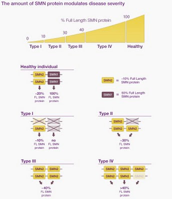
Here's an image taken from a website (yes, without permission).  It shows how the amount of SML protein (the  gene product needed for normal phenotype) depends on which alleles a person has, for two genes, SNM1 and SMN2, one of which, SMN2, is poorly expressed and may be duplicated.

Unfortunately the figure is not very clear (I think it must have been prepared for a different context, one with some explanatory text).  The first two genotypes are OK - the healthy individual is shown as having two functional copies of SML1 and of SML2, and the Type I as having two defective copies of SML1 and one defective and one functional copies of SML2.  But  the phenotypes don't make sense.  The amount of SML1 protein in a normal person is shown as 100%, but the amount of SML2 protein is shown as 20%.  100% of what, and 20% of what?  If the SML1 level is 100% of normal SML1 protein, then the SML2 protein level in a normal person should be 100% too.  If we instead think they might mean 100% of total (SML1 + SML2) protein, then the total SML1 protein can't be 100% because there's also a contribution from SML2 (the total would have to be 120%).

It gets more confusing when we look at the Type II individual.  Here the defective allele of SML1 is shown aligned with and thus allelic to a functional copy of SML2.  Well, maybe this genotype arose by a gene conversion event that replaced one SML1 allele with a paralogous SML2 allele...  Indeed, when I looked at a pdf about SMA genetics, written for parents, I found statements that gene conversion is frequent and that many patients have replaced one or both copies of the SML1 gene withSML2.

While I've been writing this the moderator of the session has been showing us how to collect and insert a reference list into a Wordpress blog.  I wasn't paying enough attention to see how he did this - he used a Wordpress plugin, but there was quite a bit of muttering about the site being slow.

Now he's going to show us another plugin.  e-Pub???  The text on the screen is just too small to read - he embiggened it once, but it seems to have shrunk back.  He says ePub is nicer than pdfs, but I've no idea what it does or where to find the plugin.